Esophagus, Stomach & Upper GI · Article 22
How Can Advanced Endoscopy Detect Early Malignant and Premalignant Lesions?
Advanced endoscopy malignancy requires a structured, evidence-based clinical approach. This review summarizes the key diagnostic, management, monitoring and decision points for gastroenterology and hepatology practice.
Advanced endoscopy malignancy: practical clinical approach
This page focuses on Advanced endoscopy malignancy with emphasis on decisions that change patient care, common pitfalls, and the current evidence base.
External guidance for Advanced endoscopy malignancy
Clinical takeaway: For Advanced endoscopy malignancy, decisions should integrate current guidelines, patient-specific risk, and the evidence summarized below.
Detection comes before technology
The most important ‘advanced’ skill is systematic inspection. Missed lesions frequently reflect poor preparation, inadequate distension, folds not examined, insufficient time or failure to recognize subtle morphology. Technology amplifies attention; it does not create it.
Image enhancement reveals pattern
Virtual chromoendoscopy highlights vascular and pit-pattern abnormalities. Dye can accentuate topography and lesion borders. Magnification can further characterize pit and microvascular architecture in experienced hands. These methods are useful in Barrett’s esophagus, gastric premalignant disease, IBD surveillance and colorectal lesions.
Characterization determines the resection plan
The endoscopist must estimate lesion boundaries, morphology and likelihood of deep submucosal invasion. Features suspicious for deep invasion may favor surgery or other oncologic management rather than piecemeal endoscopic resection. Lesions that appear superficial can often be treated endoscopically.
EMR versus ESD
EMR is efficient and widely used for many superficial lesions, especially colorectal lesions amenable to complete resection. ESD enables en bloc removal of larger or fibrotic lesions and yields superior pathologic margin assessment, but it is technically demanding and carries greater procedure time and complication risk. Referral patterns should reflect local expertise.
Pathology is part of the procedure
A technically complete resection is not the end of management. En bloc specimen orientation, depth of invasion, lymphovascular invasion, differentiation and margins determine whether endoscopic therapy was curative or whether surgery/oncology evaluation is still required.
A practical clinical approach
- Identify high-risk patients and use high-quality preparation.
- Inspect systematically with HD white light before enhancement.
- Use virtual/dye chromoendoscopy to define subtle surface and vascular abnormalities.
- Estimate invasion depth and lesion boundaries before biopsy or resection decisions.
- Choose EMR, ESD or surgery based on lesion characteristics and expertise.
- Review pathology for curative criteria and establish surveillance.
Common errors to avoid
- Biopsying a large superficial lesion repeatedly and creating fibrosis before referral for definitive resection.
- Using piecemeal resection when en bloc pathology is crucial and ESD expertise is available.
- Equating visual disappearance with oncologic cure.
- Ignoring procedure quality because advanced imaging is available.
What should trainees remember?
Advanced endoscopy works as a chain: detect, characterize, stage, resect, interpret pathology and surveil. Failure at any link can erase the benefit of sophisticated imaging.
Frequently asked questions
Can early GI cancers be cured endoscopically?
Selected superficial neoplasms can be curatively resected endoscopically when invasion and pathology criteria are favorable.
What is the difference between EMR and ESD?
EMR is generally simpler and often piecemeal for larger lesions; ESD permits en bloc resection of larger lesions but requires advanced expertise.
Does enhanced imaging replace pathology?
No.
Suggested free reading
Continue with these free books by Dr. Alan B. R. Thomson:
References
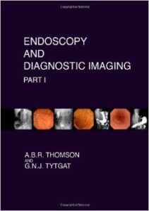
1. Thomson ABR. Endoscopy and Diagnostic Imaging, Parts I-II. CAPstone Academic Publishers; 2012. ISBN 978-1477400579 and 978-1477400654.
2. Thomson ABR. Images in Gastroenterology and Hepatology, Parts 1-2. CAPstone Academic Publishers; 2021. ISBN 979-8719829074 and 979-8743669325.
3. Morgan DR, et al. ACG Clinical Guideline: Gastric Premalignant Conditions. 2025.
4. Shaheen NJ, et al. Updated ACG Guideline: Barrett’s Esophagus. Am J Gastroenterol. 2022;117:559-587.
5. Rex DK, et al. Quality Indicators for Colonoscopy. ACG/ASGE Position Statement. 2024.
Free GI & Hepatology Updates
Continue Learning With Dr Alan B. R. Thomson
Receive evidence-based clinical teaching, updated reviews, practical reference material and new free GI and hepatology resources.
Free to subscribe. Educational resources on GIandHepatology.com remain freely available without registration.